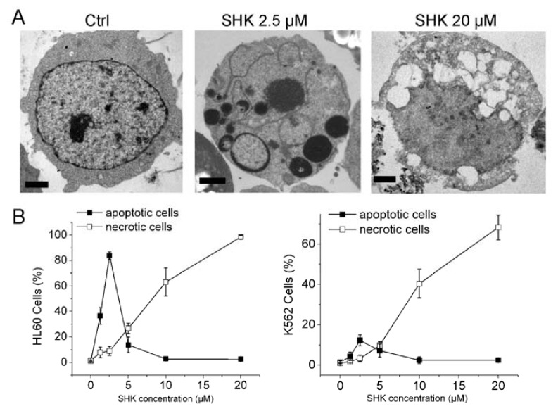
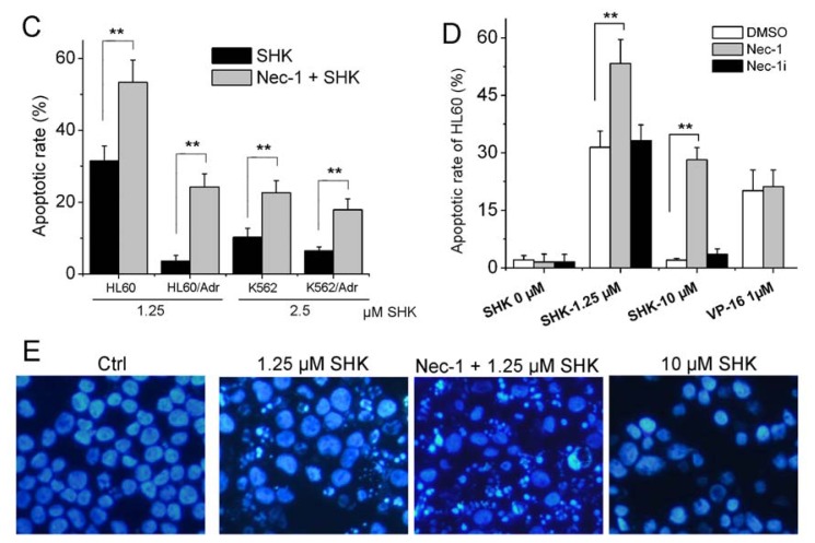
Figure 1.
Nec-1 enhances shikonin-induced apoptosis in leukemia cells. (A) K562 cells were treated with various concentrations of shikonin for 12 h. Transmission electron micrograph showing that shikonin induced a typical apoptotic morphology at 2.5 μM, and a feature of necroptosis at 20 μM. Bar = 2 μm; (B) Cells were incubated with varying concentrations of shikonin for 12 h. Total cell death was measured by Vital dye exclusion assay and Hoechst-staining; (C) HL60, HL60/Adr, K562 and K562/Adr cells were treated with 1.25 or 2.5 μM shikonin for 12 h in the absence or presence of 60 μM Nec-1. Cells apoptotic rate was determined as described in Materials and Methods; (D) HL60 cells were treated with 1.25, 10 μM shikonin, or 1 μM VP-16 for 12 h in the absence or presence of 60 μM Nec-1 or Nec-1i. Cells apoptotic rate was determined as described in Materials and Methods. (E) Primary leukemia cells were treated with shikonin for 12 h in the presence or absence of Nec-1, and nuclei were stained by hoechst. Data are mean ± SD or representative of at least three independent experiments, and analyzed by Student’s t test. ** p < 0.01 compared with SHK treated group.


