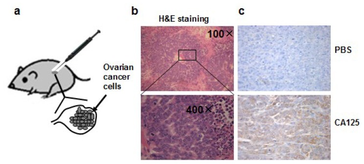
Figure 1.
(a) Orthotopic ovarian tumor model in this study; (b) H&E staining of tumor section showing tumor cells and surrounding tissue; (c) Expression of ovarian cancer biomarker CA125 detected by immunohistochemical staining. Upper panel: section was incubated with PBS buffer instead of CA125 primary antibody for immunohistochemical staining (200× magnifications), bottom panel: section incubated with CA125 antibody.

