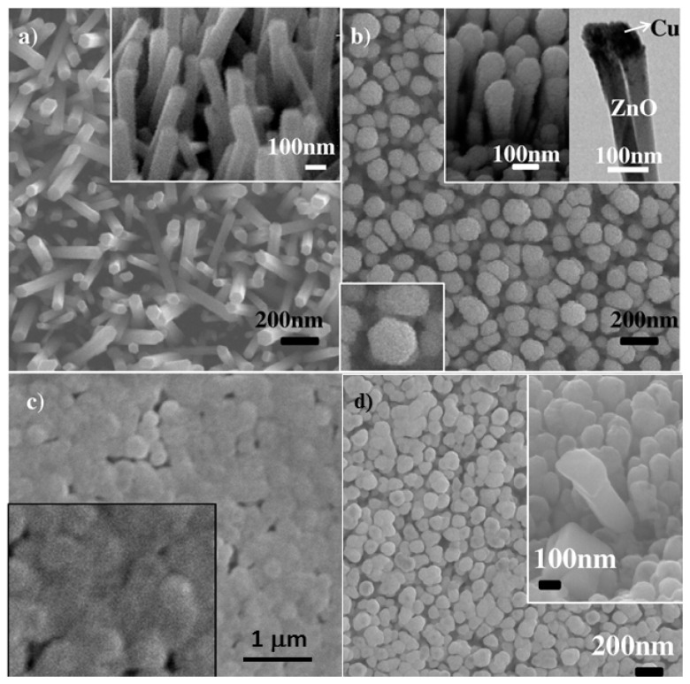
Figure 19.
Cu film oxidation on ZnO nanowire arrays: Top view SEM images (a) ZnO nanorods grown by hydrothermal method, inset: a 30° tilt view of nanorods; (b) ZnO-Cu core-shell nanorods after deposition of Cu and upper right inset (left) showing their nail-shaped structure, upper right inset (right): TEM image of ZnO-Cu core-shell nanorods; bottom left inset: zoom in view of a hexagon shaped nanorod tip; (c) ZnO-Cu core-shell nanorods annealed at 400 °C in ambient air for 30 min, inset: zoom in view of “flat” and continuous CuO film bridging adjacent ZnO nanrods; (d) Top view SEM image of ZnO-CuO core-shell nanowire arrays: 80 sccm oxygen flow rate and 100 mabr pressure.

