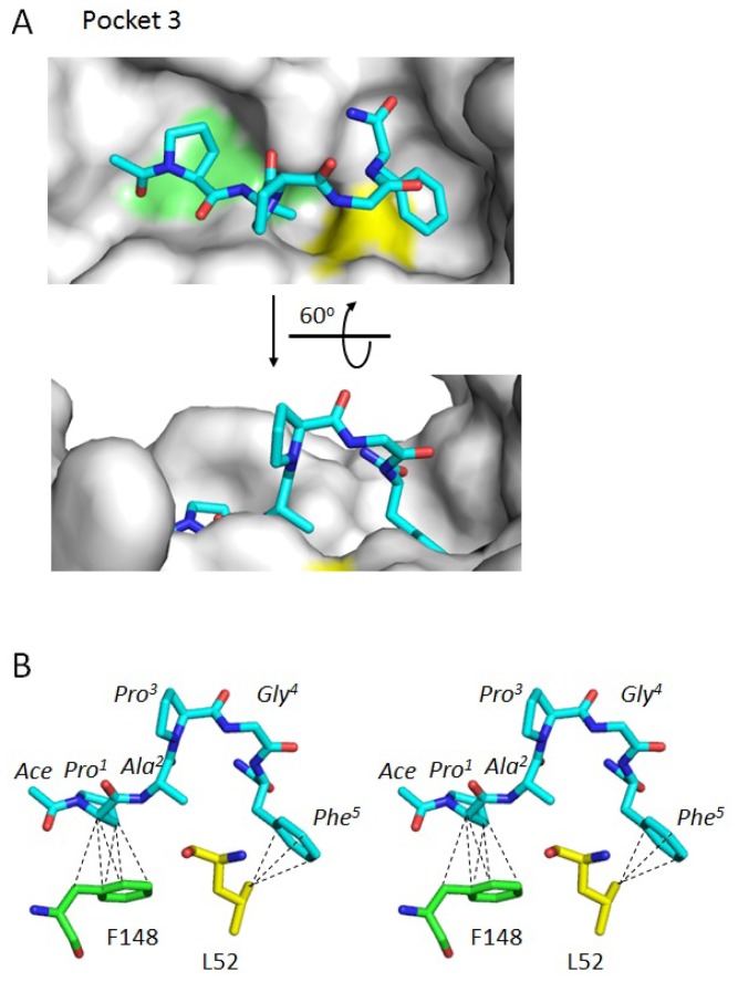
Figure 6.
Docking simulation. (A) Residues forming Pocket 3 and its vicinity are shown by surface representation in gray except for F148 in light green and L52 in yellow. The type 2 motif peptide is represented by a stick model in which atoms of carbon, nitrogen and oxygen are shown in cyan, blue and red, respectively, in the upper panel. A side view of the structure shown in the upper panel obtained by rotating 60° around the indicated axis is presented in the lower panel; (B) The peptide shown in (A) upper panel is rotated 75° around the axis and represented in stereoview (parallel viewing method). Hydrophobic interactions between the type 2 motif peptide and ALG-2 are shown for F148 (carbon atoms, green) and L52 (carbon atoms, yellow). Broken lines indicate hydrophobic interactions with distances shorter than or equal to 3.9 Å (see Table 3 for details).

