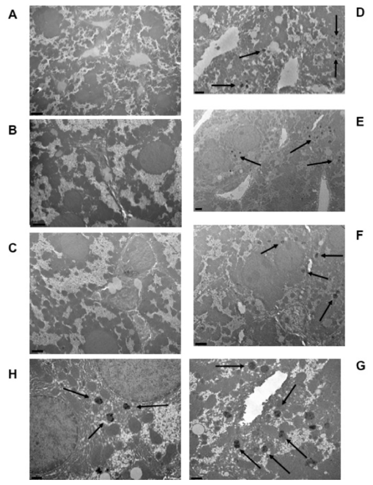
Figure 2.
Metal deposits in liver cells of wild-type mice after 7 months of exposure to an aimara-containing diet. Transmission electron microscopy was performed on the liver from control mice (A–C) and aimara-fed mice (D–H). Scale bars appear in the left bottom corners of photos and represent 5 μm (A), 2 μm (B–F) and 1 μm (G and H). Black arrows indicate the metal deposits.

