Figure 7.
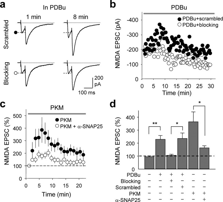
SNAP-25 is required for PKC-induced synaptic insertion of NMDARs in hippocampal slices. a–d, Synaptic NMDA currents (NMDA EPSCs) were recorded from CA3 pyramidal neurons in the presence of CNQX, LY303070, and picrotoxin (to block kainate receptors, AMPARs and GABAA receptors), and 1 mm Mg2+ (to reduce Mg2+ block) at Vh = −40 mV. Mossy fibers were activated by paired-pulse electrical stimulation in the dentate gyrus. a, b, Representative traces (a) and time course data (b) showing that application of the PKC-activating phorbol ester PDBu (1 μm, delivered via the recording pipette solution) potentiated NMDA EPSCs in the presence of the scrambled peptide (as in Fig. 6). Loading the postsynaptic cell with the SNAP-25 blocking peptide abolished PKC potentiation. b, Representative time course data showing block of PKC potentiation by blocking peptide (○), but not by scrambled peptide (•). c, An anti-SNAP-25-antibody greatly reduced PKC-induced potentiation of NMDA EPSC. Summary time course showing that the catalytic subunit of PKC, PKM (1 μm, delivered via the patch pipette), potentiated NMDA EPSC; a function-blocking anti-SNAP-25 antibody (α-SNAP-25) inhibited PKM potentiation. Each data point was the average of 6–8 experiments in which responses were binned every minute. d, Summary of data in a–c. All data are expressed as means ± SEMs of percent control response at break in. *p < 0.05; **p < 0.01.

