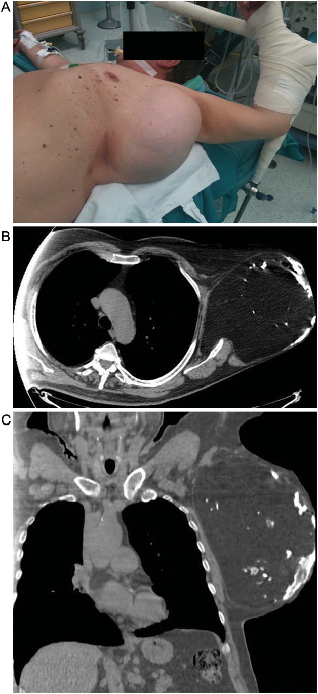
Figure 1:

(A) Preoperative appearance of the chest wall mass. (B–C) Chest CT scan evidenced a solid neoplasm measuring 27 cm in its major axis, apparently originating from the left serratus anterior muscle. The mass showed a homogeneous fat density with spotted areas of calcification. No direct signs of chest wall invasion were detected.
