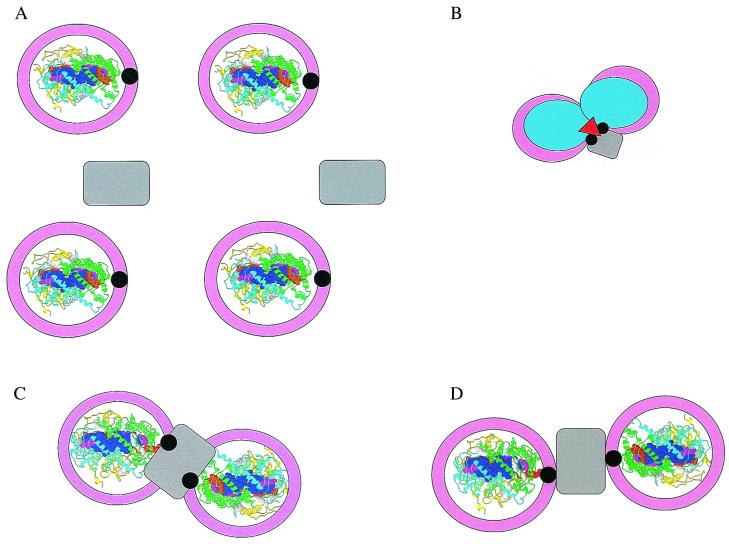
Figure 1.
(A) Model of the core complex in which each RC (Rasmol structure of the Rb. sphaeroides RC, Protein Data Bank (RCSB) accession no. 1aij) is surrounded by a closed ring of LH1 (pink) interrupted by PufX (black) and interacting with the RC at the QB site (orange space-fill tail of QB). The bc1 complex (gray) is elsewhere in the membrane. In the Rasmol presentation, the RC is viewed from the extracellular side of the membrane, the H-polypeptide is yellow, the L-polypeptide is cyan, the M-polypeptide is green, bacteriochlorophyll are blue, bacteriopheophytin are magenta, and QA and QB are orange. (B) Model of an intimate RC dimer (light blue) in which PufX and another possible component (red) link LH1 with the RC and cytochrome b (28, 29). Other color assignments are as for A. (C) Model of an RC-cytochrome b-RC complex in which two RC are intimately associated with a bc1 complex linked by PufX (23). Colors are as in A. (D) Model of a core complex-cytochrome b-core complex in which the linkage to cytochrome b is through PufX (22). Colors are as in A.

