FIGURE 3.
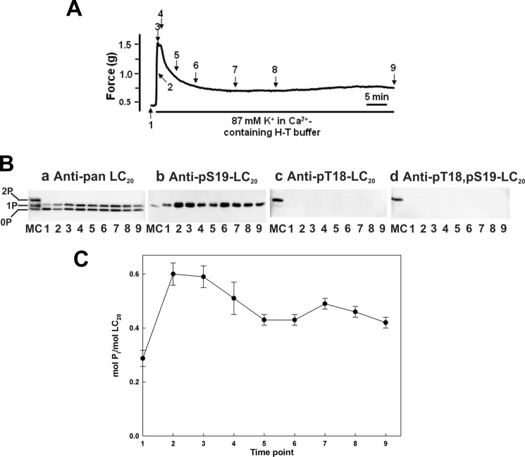
Contraction and LC20 phosphorylation in intact rat caudal arterial smooth muscle in response to KCl-induced depolarization in the presence of Ca2+. A, intact rat caudal arterial smooth muscle strips were treated with 87 mm KCl in Ca2+-containing H-T buffer and the contractile response was recorded. Separate tissues were harvested at the indicated times during the contraction for analysis of LC20 phosphorylation by Phos-tag SDS-PAGE and Western blotting with antibodies to LC20 (panel a), Ser(P)19-LC20 (panel b), Thr(P)18-LC20 (panel c), and Thr(P)18,Ser(P)19-LC20 (panel d). Numbers below the gel lanes correspond to the time points in A. MC denotes control tissue (Triton-skinned rat caudal arterial smooth muscle treated with microcystin at pCa 9 for 60 min) to identify unphosphorylated, mono-, and diphosphorylated LC20 bands. C, cumulative quantitative data showing the time course of LC20 phosphorylation stoichiometry; as shown in panel b, phosphorylation occurred exclusively at Ser19 under these conditions. Values represent the mean ± S.E. (n = 3).

