FIGURE 2.
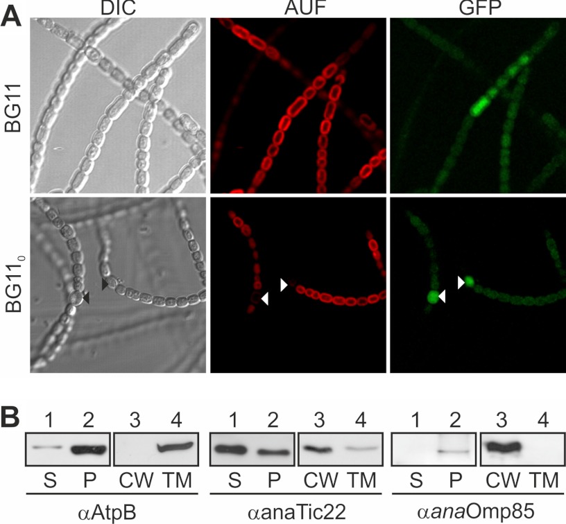
The localization of anaTic22. A, light microscopy images of Anabaena sp. mutant strain AFS-PDGF-tic22 (left) grown in BG11 (top) or BG110 (bottom), and confocal fluorescence images of the chlorophyll autofluorescence (middle) and GFP (right) are shown. Triangles mark heterocysts. B, Anabaena sp. was fractionated into soluble (S) and pellet fraction (P); the latter in cell wall (CW) and thylakoid membranes (TM). Samples were immunodecorated with indicated antibodies. AUF, autofluorescence, DIC, differential interference contrast.

