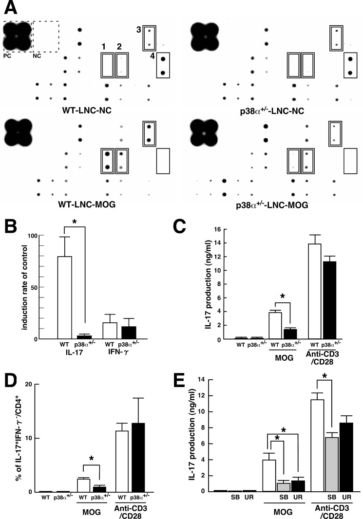
FIGURE 3.
Up-regulation of IL-17 in antigen-specific LNC mediated by p38α. A, Western blot array analysis of cytokines from cultured LNC stimulated with MOG(35–55). LNC from WT and p38α+/− mice at 8 dpi were stimulated in vitro with MOG(35–55) as an antigen-specific response. The supernatants were subjected to Mouse Cytokine Antibody Array 2. Compared with the unstimulated control, MOG(35–55)-induced and -reduced molecules are indicated by a double-line and a single-line square, respectively. Each number on the membrane represents the following: 1, IL-17; 2, IFN-γ; 3, IL-3; and 4, MCP-1. Positive and negative controls on the membrane in the reaction are indicated by a dotted square. B, change in expression of IL-17 and IFN-γ observed in Western blot array analyzed by densitometer. Each bar (WT, white bars; p38α+/−, black bars) is expressed as a percentage of the individual control signal without MOG(35–55) stimulation. Data are shown as mean ± S.E. (n = 6). The difference between the two groups was statistically significant (*, p < 0.05) as determined by Student's t test for unpaired values. C, MOG(35–55)-induced IL-17 in LNC. LNC from WT (white bars) and p38α+/− (black bars) mice at 8 dpi were stimulated in vitro with MOG(35–55) as an antigen-specific response or anti-CD3/CD28 antibodies as an APC-free response. The resulting supernatants were subjected to ELISA for IL-17. Data are shown as means ± S.E. (n = 4). The difference between the two groups was statistically significant (*, p < 0.05) as determined by Student's t test for unpaired values. D, detection of IL-17-producing CD4 T cells by FACScan. LNC from WT (white bars) and p38α+/− (black bars) mice at 8 dpi were stimulated in vitro with MOG(35–55) or anti-CD3/CD28 antibodies and subjected to surface staining for CD4 and intracellular staining for IL-17 and IFN-γ. Quantification of CD4+IL-17+IFN-γ− T cells was performed with FlowJo software. Data are shown as mean ± S.E. (n = 4). The difference between the two groups was statistically significant (*, p < 0.05) as determined by Student's t test for unpaired values. E, effects of p38 inhibitors on IL-17 production in LNC. LNC from WT mice at 8 dpi were stimulated in vitro with MOG(35–55) or anti-CD3/CD28 antibodies in the absence or presence of p38 inhibitor (SB203580 (SB), shaded bars; UR-5269 (UR), black bars). The resulting supernatants were subjected to ELISA for IL-17. Data are shown as mean ± S.E. (n = 3). *, p < 0.05, significantly different from the value without p38 inhibitors (white bars) (ANOVA with Bonferroni method).

