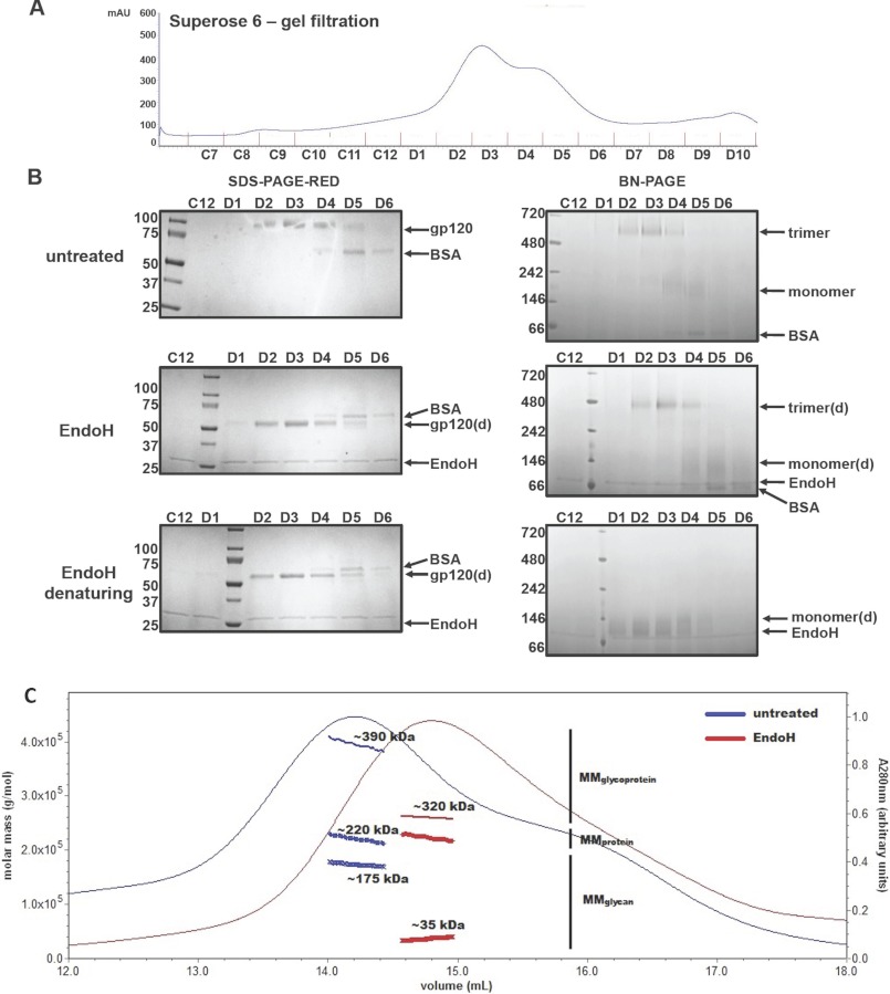
FIGURE 6.
Biophysical analysis of SOSIP.664GnTl−/− trimers incubated with or without Endo H. A, untreated SOSIP.664GnTl−/− trimers were fractionated using a Superose-6 column. The fractions were treated with or without Endo H and analyzed by reducing SDS-PAGE (B, left panels) or BN-PAGE (B, right panels). B, top panels, untreated trimers; middle panels, Endo H-treated trimers (3 h at 37 °C, pH 5.5); bottom panels, denatured and Endo H-treated trimers. The species associated with each band are indicated. C, overlay of the A280 UV absorbance traces from Superose-6 column fractionations of untreated (blue) and Endo H-treated (3 h at 37 °C, pH 5.5; red) SOSIP.664GnTl−/− trimers. UV/MALS/RI signals were recorded as the samples eluted from the column. Absolute molar masses calculated from these measurements are shown as thick blue and green connected dots under the main peaks for the samples obtained with and without Endo H treatment (see also Table 2). d, deglycosylated.

