FIGURE 9.
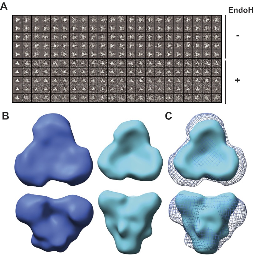
Single particle image reconstructions of negatively stained SOSIP.664GnTl−/− trimers. A, five representative, reference-free two-dimensional class averages (left images) and the corresponding images of oriented raw particles are shown. The top five class averages belong to the fully glycosylated trimers, the lower five to the Endo H-treated trimers. The white scale bar, located in the top left image, corresponds to 50 Å. B, top and side views of trimer reconstructions of untreated, fully glycosylated (blue) and Endo H-treated trimers (cyan) that have been low-pass filtered to 20-Å resolution. Contour levels corresponding to experimentally determined masses by SEC-UV/MALS/RI (see “Materials and Methods”) were calculated using the “volume” algorithm in EMAN. Untreated trimers were contoured using a volume of ∼390 kDa, whereas the Endo H-treated trimers were contoured with a volume of ∼260 kDa. The overall shapes of the trimers are conserved. C, overlay of untreated (blue mesh) and Endo H-treated (cyan surface) trimers, highlighting the conserved morphology and the size difference between the trimers.

