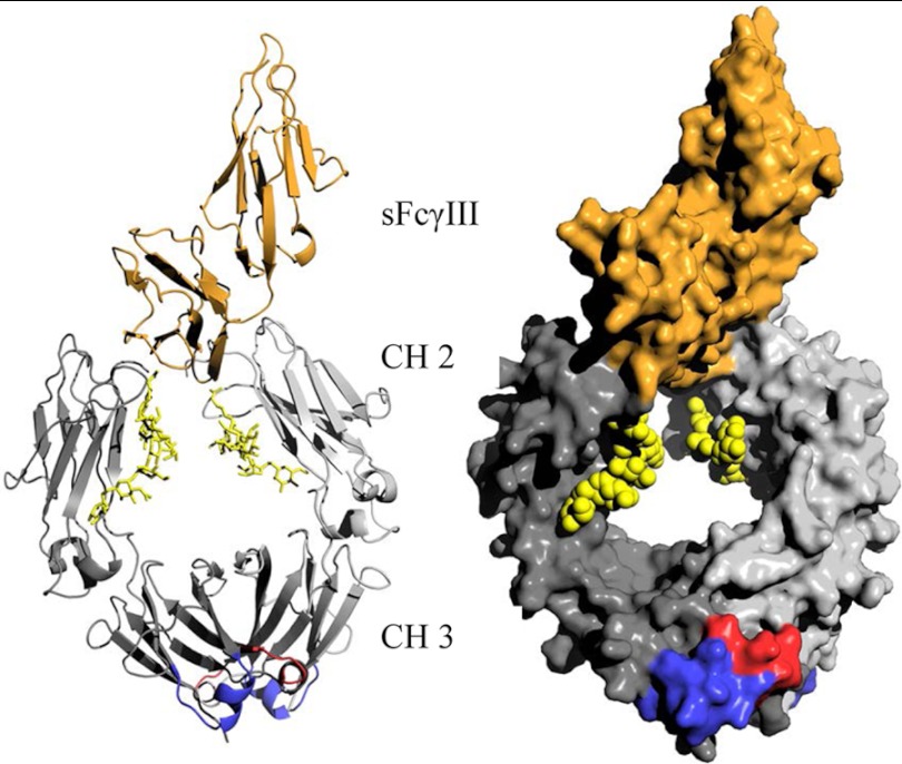
FIGURE 6.
Cartoon and surface presentation of human Fc from IgG1 (gray) in complex with soluble CD16 (sFcγIII receptor, orange). The location of the CD16 binding site is on the N-terminal side of the CH2 domain of Fc, whereas engineered antigen-binding sites in Fcabs such as the HER2/neu in H10-03-6 are located at the C-terminal part of the CH3 domain (bottom). Each of the two antigen-binding sites in the Fc homodimer (dark and bright gray indicate the two chains) is composed of residues in the AB (red) and in the EF (blue) loop (5). Sugar residues in the N-linked glycosylation sites are indicated in yellow.

