
FIGURE 3.
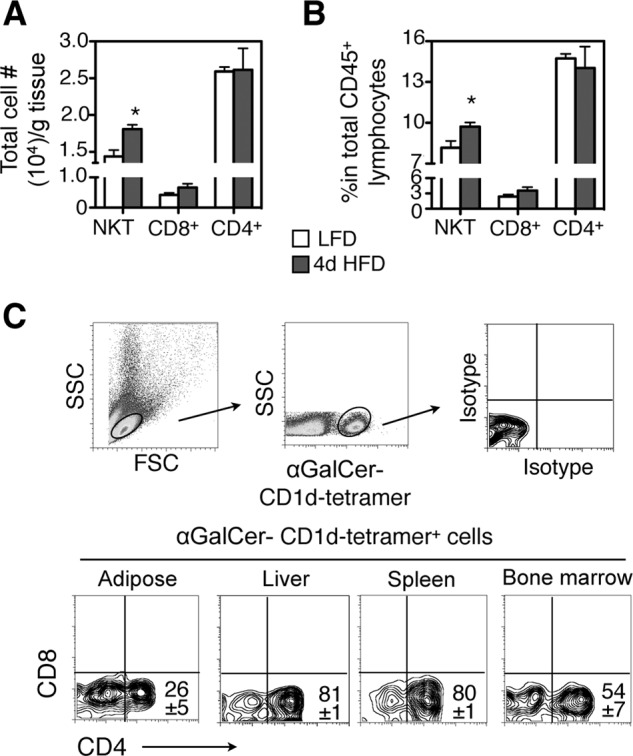
Adipose-resident type I NKT cells increase with 4d HFD and are predominantly CD4−CD8−. Analysis of NKT cells in epididymal adipose tissue of 6-week-old male lean mice. A and B, total number of NKT cells per gram of adipose tissue (A) and percent (B) of NKT cells in total adipose lymphocytes are shown. n = 12 mice with three repeats. C, characterization of cell-surface markers CD4-CD8 of αGalCer-CD1d-tetramer-positive NKT cells from indicated tissues. Numbers in the flow histogram indicate the percent of CD1d-tetramer-positive NKT cells. n = 10–12 mice with at least 4 repeats. Values represent mean ± S.E. *, p < 0.05.