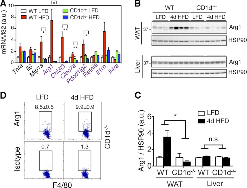
FIGURE 5.
Loss of NKT cells alters inflammatory responses in adipose tissue with 4d HFD challenge. 6-week-old WT or CD1d−/− mice were placed on LFD or HFD for 4 days. A, Q-PCR analysis of M1 and M2 (purple) genes in the WAT (n = 12–15 mice each group, three repeats). B and C, Western blot analysis of Arg1 expression in WAT (upper) and liver (lower) of WT and CD1d−/− cohorts on either LFD or 4d HFD. n = 3–4 mice each with two repeats. Quantitation shown in C. D, flow cytometric analysis of intracellular Arg1 in CD45+ F4/80+ cells of adipose tissue of CD1d−/− mice under LFD versus 4d HFD. Percent of positive cells is indicated. Values represent averages of one experiment (n = 6 mice each) with two repeats. Values represent mean ± S.E. n.s., not significant; *, p < 0.05 and **, p < 0.01 comparing the two groups or groups included in brackets.

