FIGURE 7.
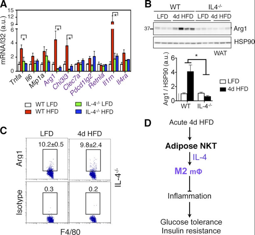
Loss of IL-4 alters inflammatory responses in adipose tissue with 4d HFD challenge. A, Q-PCR analysis of M1 and M2 (purple) genes in the WAT of 4 cohorts (n = 4–5 mice each cohort). B, Western blot analysis of Arg1 expression in WAT of WT and IL-4−/− cohorts on either LFD or 4d HFD with quantitation shown below. n = 3–4 mice each, two repeats. C, flow cytometric analysis of intracellular Arg1 in CD45+ F4/80+ cells of adipose tissue of IL-4−/− mice under LFD versus 4d HFD. Percent of positive cells is indicated. Values represent averages of one experiment (n = 6 mice each), two repeats. Values represent mean ± S.E. *, p < 0.05. D, proposed function of adipose-resident NKT cells in response to acute HFD feeding. Upon acute 4d HFD challenge, NKT cells promote M2 polarization and Arg1 expression via IL-4 in adipose tissue, thereby promoting systemic insulin sensitivity and glucose tolerance.

