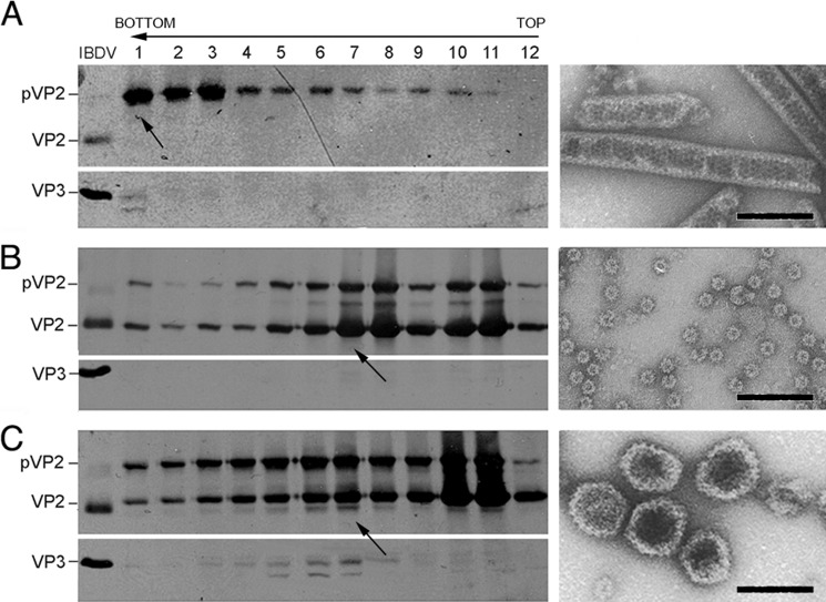
FIGURE 2.
Coexpression of pVP2-452 and IBDV polyprotein form is necessary for T = 13 VLP assembly in H5 insect cells. A, wild-type rBV/Poly assemblies were purified by ultracentrifugation on sucrose gradients, collected in 12 fractions, concentrated by ultracentrifugation, and analyzed by SDS-PAGE and Western blot analysis using anti-VP2 (left panel, top) and anti-VP3 antibodies (left panel, bottom). The direction of sedimentation was from right to left, with fraction 12 at the gradient top. A representative electron micrograph image is shown of pVP2 tubular structures from lower gradient fractions (arrow). B and C, assemblies from cells coinfected with rBV/Poly and rBV/VP2-441 (B) or rBV/Poly and rBV/pVP2-452 (C) were analyzed as in A. Representative electron micrographs (arrow) of T = 1 SVP from top fractions (B, right panel) and IBDV-like capsids from the center fractions (C, right panel). Bands are indicated for VP2 and VP3 in a purified IBDV sample (left panel). Scale bar = 100 nm.

