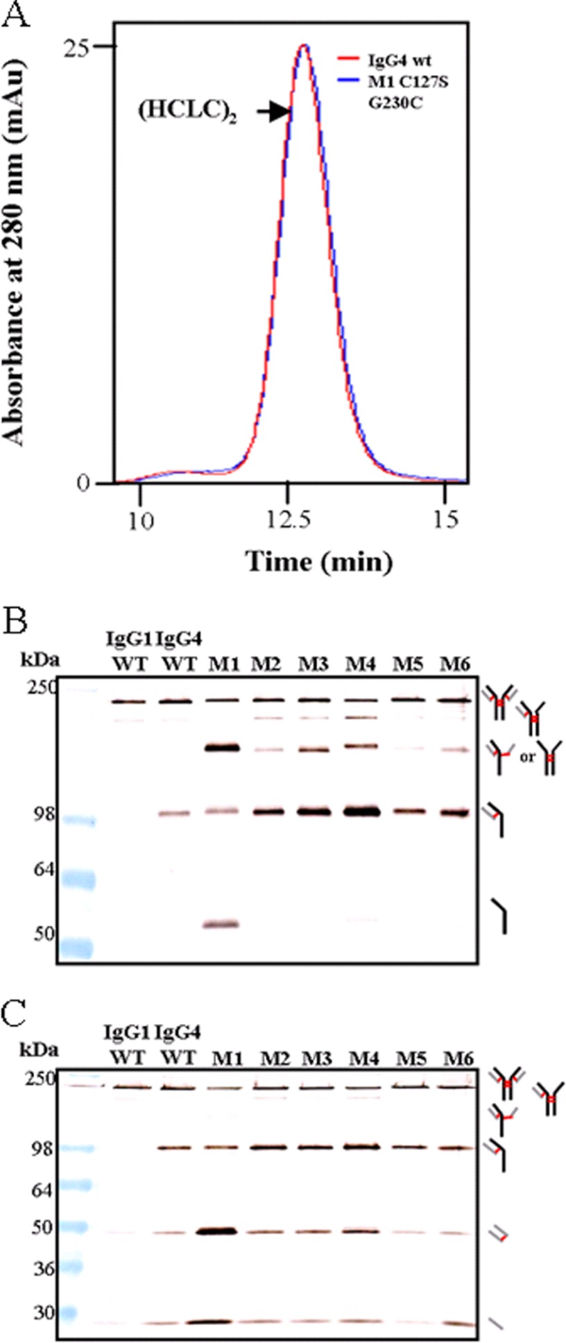
FIGURE 3.
SEC HPLC and immunoblot of IgG4 molecules with altered DSB arrangements toward an IgG1-like DSB, including mutants with a 3-residue spacer within the hinge region. All mutants include C127S in addition to the mutation noted. A, SEC HPLC trace comparison of IgG4 WT and M1 (C127S/G230C). B, comparison of IgG4 WT with mutants that had a Cys introduced at different positions in the CH1. Samples were run under non-reducing conditions and probed with an anti-human Fc antibody. C, comparison of the same samples, also run under non-reducing conditions but probed with an anti-human κ antibody. For both A and B, thick black lines indicate HC, thick gray lines indicate LC, and red lines indicate DSBs.

