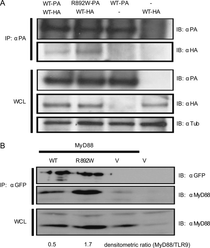
FIGURE 3.
TLR9 R892W co-immunoprecipitates with WT TLR9, binds to MyD88, but fails to reach CpG DNA containing endosomes. A, HEK293T cells were transiently co-transfected with TLR9 WT-protein A (PA) and TLR9 R892W-HA fusion protein expression vectors and lysed after 48 h. Cleared lysates were immunoprecipitated (IP) with α-protein A antibody and immunoblotted (IB) for protein A or HA. Whole cell lysates were immunoblotted for protein A, HA, and tubulin. One of two representative experiments is shown. B, HEK293T cells were transfected with TLR9-YFP (WT), TLR9 R892W-YFP (R892W), or empty pEYFP vector (V) together with MyD88-AU1 (MyD88) as indicated. The lysates were immunoprecipitated for GFP (reacts with YFP) and immunoblotted for GFP and MyD88. Whole cell lysates (WCL) were immunoblotted for MyD88. Note that endogenous MyD88 is detected at low abundance in all lanes. TLR9 and MyD88 bands were subjected to densitometry analysis using ImageJ. One of three independent experiments shown.

