FIGURE 3.
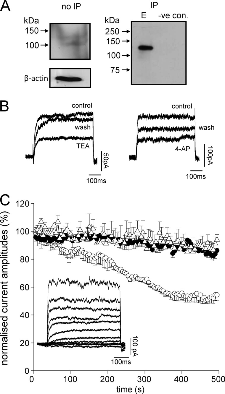
DAOY cells express functional Kv2.1 channels. A, Western blots showing the presence of Kv2.1 channel protein in DAOY total cell lysate. The left-hand blot shows detection of Kv2.1 using an anti-Kv2.1 antibody in total DAOY cell lysate without immunoprecipitation (no IP); the right-hand blot is from lysate following immunoprecipitation with anti- Kv2.1 antibody (IP; E, elution). The lane labeled negative control was performed exactly as the IP but omitting the anti-Kv2.1 antibody. B, left: example voltage-gated K+ currents evoked in DAOY cells by step depolarizations to +50 mV before (control) during (TEA) and after (wash) exposure to 5 mm TEA. Right: example K+ currents evoked by step depolarizations to +50 mV before (control) during (4-AP) and after (wash) exposure to 10 mm 4-AP. C, normalized mean time courses (± S.E. bars) of K+ current amplitudes evoked by successive step depolarizations from −90 to +50 mV (100 ms). Cells were dialyzed with a pipette solution containing either no antibody (control, solid circles, n = 6 cells), an anti-Kv2.1 antibody (0.5 μg/ml, open circles, n = 6 cells) or an anti-Kv4.3 antibody (0.5 μg/ml, open triangles, n = 6 cells). Currents became significantly reduced (p < 0.05, unpaired t test) in the presence of anti-Kv2.1 antibody as compared with controls after 200 s dialysis. The inset shows an example of a family of currents evoked in response to voltage steps from −100 to +80 mV in 20 mV increments.

