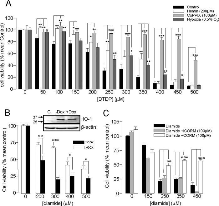
FIGURE 5.
HO-1 induction protects DAOY cells against oxidative stress. A, bar graph showing the mean (± S.E. bars) effect of oxidative stress (DTDP exposure, 0–500 μm) on DAOY cell viability. Cells were either untreated (control) or had HO-1 expression induced following incubation with hemin (200 μm, 24 h), CoPPIX (100 μm, 24 h), or hypoxia (0.5% O2, 16 h), as indicated. B, bar graph showing the mean effect on viability of diamide in DAOY cells stably expressing doxycycline-inducible HO-1 targeting shRNA. Cells were pre-exposed to hemin (200 μm, 48 h) with or without doxycycline (2 μg/ml), as indicated. The inset shows Western blot of HO-1 levels in control cells and in cells pre-exposed to hemin (200 μm, 48h) with or without doxycycline (2 μg/ml). C, bar graph showing the mean effect on viability of DAOY cells treated with diamide with and without the subsequent incubation of CORM-2 (100 μm, 3h) or iCORM (100 μm, 3 h). In A–C, each experiment was repeated three times in triplicate, and the mean number obtained each time was used for statistical analysis. (*, p < 0.05; **, p < 0.01, ***, p < 0.001; 2 way ANOVA with Bonferroni multiple-comparison test).

