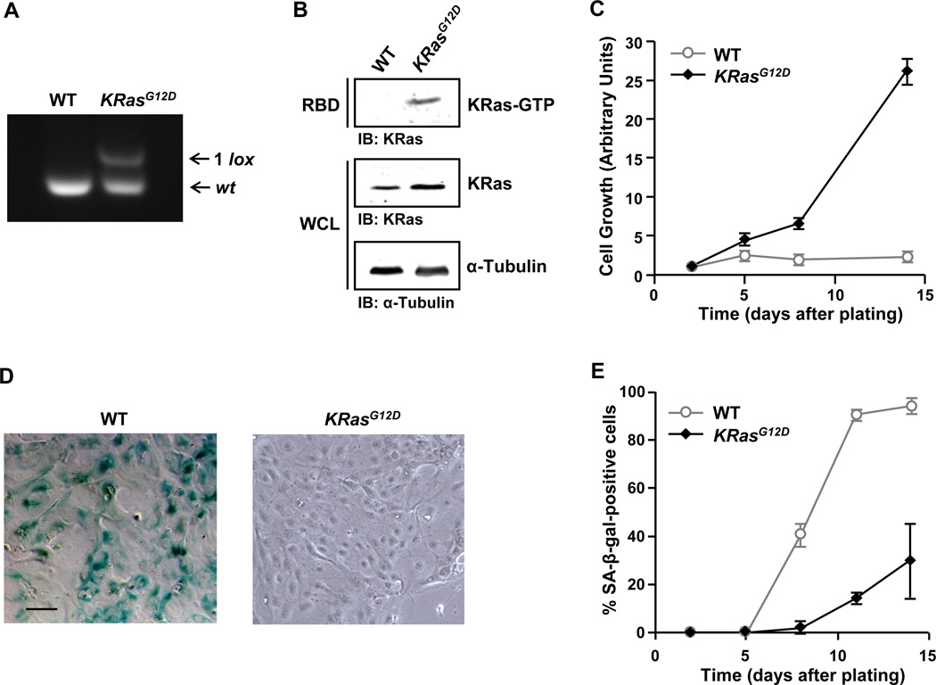
Figure 1. Oncogenic KRas protects PDEC from undergoing premature senescence.
(A) PCR analysis of genomic DNA prepared from LSL-KRasG12D PDEC infected with adenoviral-GFP (WT) or adenoviral-Cre (KRasG12D). The excision-recombination event of the LSL cassette leaves behind a single LoxP (1 lox) site.
(B) Measurement of Ras activation in WT and KRasG12D PDEC by GST-RBD pull-down assay. α-Tubulin serves as a loading control. IB, immunoblot; WCL, whole cell lysates.
(C) Growth analysis of WT and KRasG12D PDEC. The number of DAPI-stained nuclei was counted in 9 random fields of view (FOV) each containing at least 50 cells at day 2, 5, 8, and 14. The average number of nuclei present in the FOV at each time point was then normalized to the average number of nuclei per FOV at day 2. Error bars indicate standard deviation (SD). Data are representative of five independent experiments.
(D) SA-β-gal staining of WT and KRasG12D PDEC cultures at day 8. Scale bar: 100 µm.
(E) Quantification of SA-β-gal staining in WT and KRasG12D PDEC cultures at day 2, 5, 8, 11, and 14. Cells were counterstained with Hoechst 33342 for β-gal quantification. Error bars indicate SD (n = 6 FOV). Data are representative of five independent experiments. See also Figure S1.

