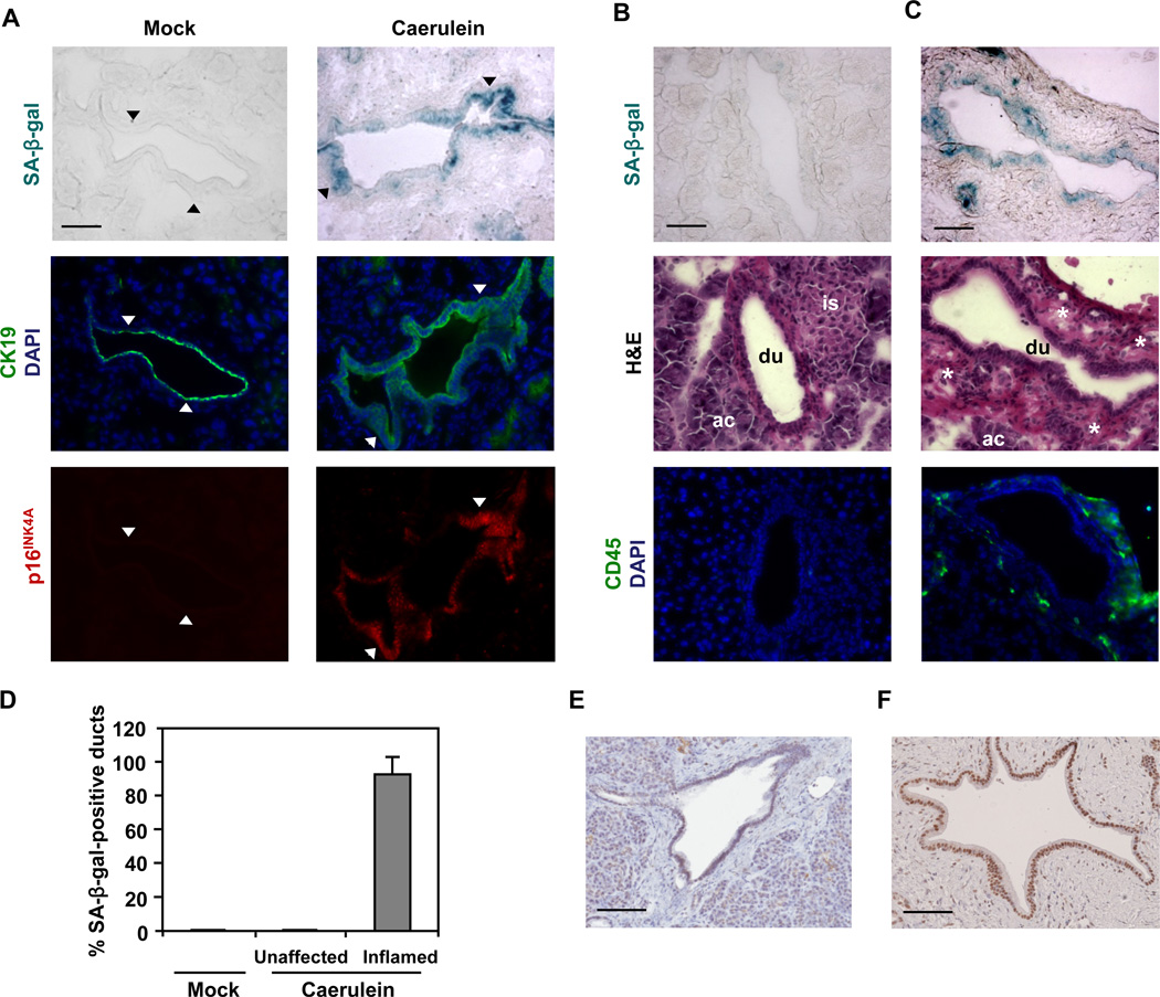
Figure 4. Inflammatory insult triggers premature senescence in pancreatic ductal epithelium in vivo.
(A–D) Caerulein was administered to 1-month-old mice as 8 hourly intraperitoneal injections (50 ng/g of body weight/injection) for two days. Three days after the last caerulein injection, pancreata were harvested. At least 3 sections were analyzed per animal (n = 5 per genotype per treatment).
(A) SA-β-gal staining and immunofluorescence staining for p16INK4A and CK19 on consecutive sections of pancreata from WT mice treated with caerulein or saline (mock). CK19 was used to identify ductal cells. Nuclei were counterstained with DAPI. Arrowheads mark corresponding areas.
(B and C) SA-β-gal staining, hematoxylin and eosin (H&E) staining, and immunofluorescence staining for CD45 on consecutive sections of pancreata from WT mice treated with caerulein. Pancreatic ducts in unaffected (B) or inflamed (C) areas are from the same tissue section. CD45 was used to identify leukocytes. Nuclei were counterstained with DAPI. ac, acinus; du, duct; is, islet; asterisk, immune infiltrate.
(D) Quantification of SA-β-gal staining in pancreata from WT mice treated with caerulein or saline (mock). Pancreatic ducts bearing 10% or more β-gal-positive cells in each duct were scored as positive. At least four pancreatic ducts were scored in each mouse. Error bars indicate SD (n = 5).
(E and F) Immunohistochemical analysis of p16INK4A in human chronic pancreatitis tissue samples. Representative areas of normal pancreatic (E) and pancreatitis (F) tissues are shown. Scale bars: 50 µm (A, B, and C), 100 µm (E and F). See also Figure S3.

