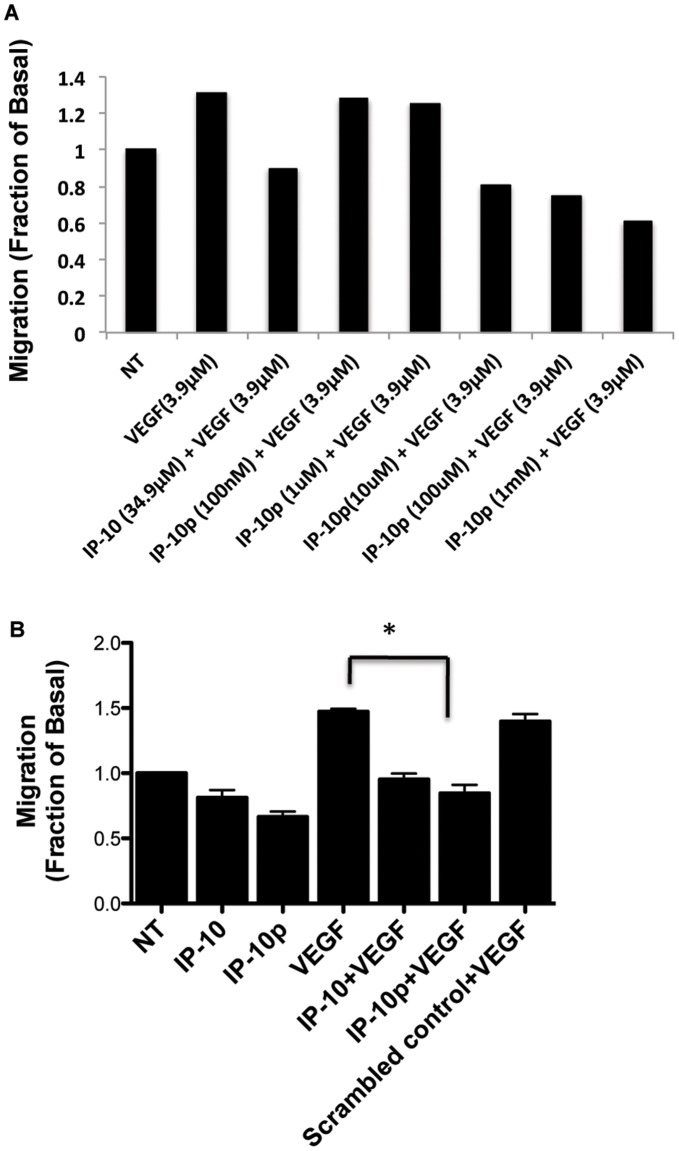
Figure 3. 2D scratch assay of stimulated HMEC-1 cells was used to analyze migration patterns.
A) The dose response used to determine the optimal concentration IP-10p (10 µM) used to compare to IP-10 (34.9 µM) B) HMEC-1 cells were grown to 80 to 85% confluence in a 12-well plate and quiesced in 0.5% dialyzed fetal bovine serum for 24 hours. A 1-mm scratch was made to the confluent of monolayer using a rubber policeman. The cells were then incubated in 0.5% dialyzed with/without IP-10 (23.2 µM), IP-10p (10 µM), VEGF (3.9 µM) or and/or scrambled control (10 µM) for 24 hours. As expected, IP-10p inhibited motility of the HMEC-1 as wells as inhibited VEGF induced motility. The results are N = 6 (average ±SEM). *P<0.05.

