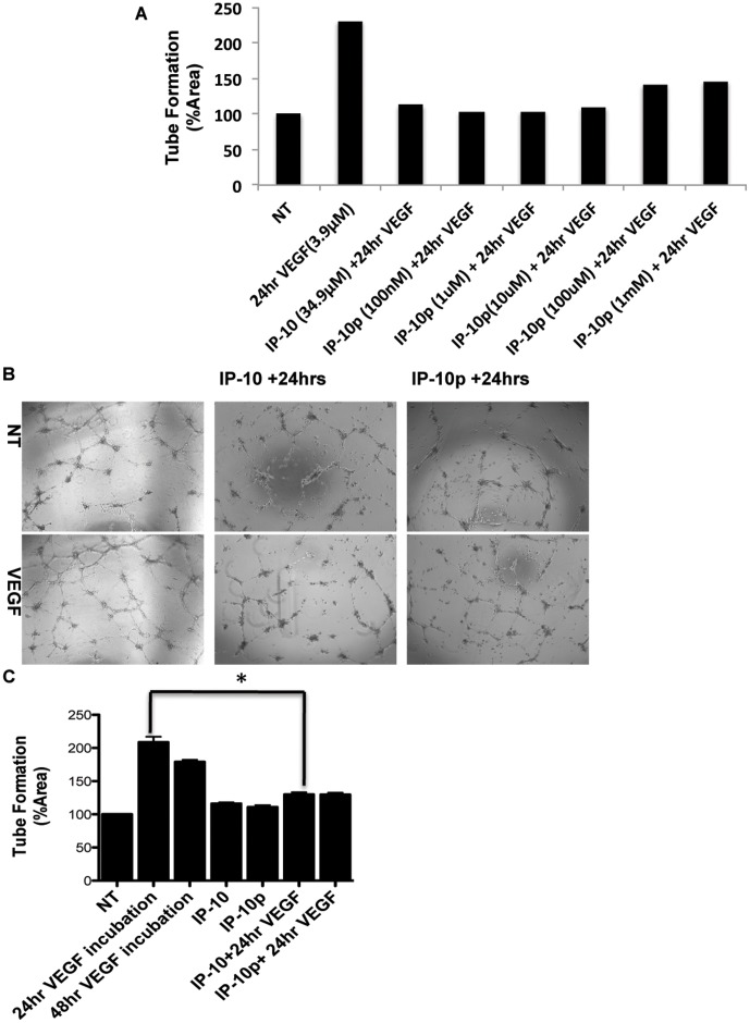
Figure 5. IP-10p induces dissociation of newly formed tubes.
A) The dose response used to determine the optimal concentration IP-10p (10 µM) used to compare to IP-10 (34.9 µM). B) HMEC-1 cells were treated with VEGF (3.9 µM) and plated on GFR-Matrigel to form endothelial tubes. The newly formed tubes were incubated in 0.5% dialyzed FBS medium for 24 hours with VEGF (48 hours) in the presence of IP-10 (34.9 µM) (24 hrs+IP-10), or IP-10p (10 µM) (24 hrs+IP-10). C) Quantification of the endothelial tube area was determined, using MetaMorph. Data shown are of at least N = 6 and normalized to no treatment (average ±SEM). *P<0.05. Original magnifications, 4X.

