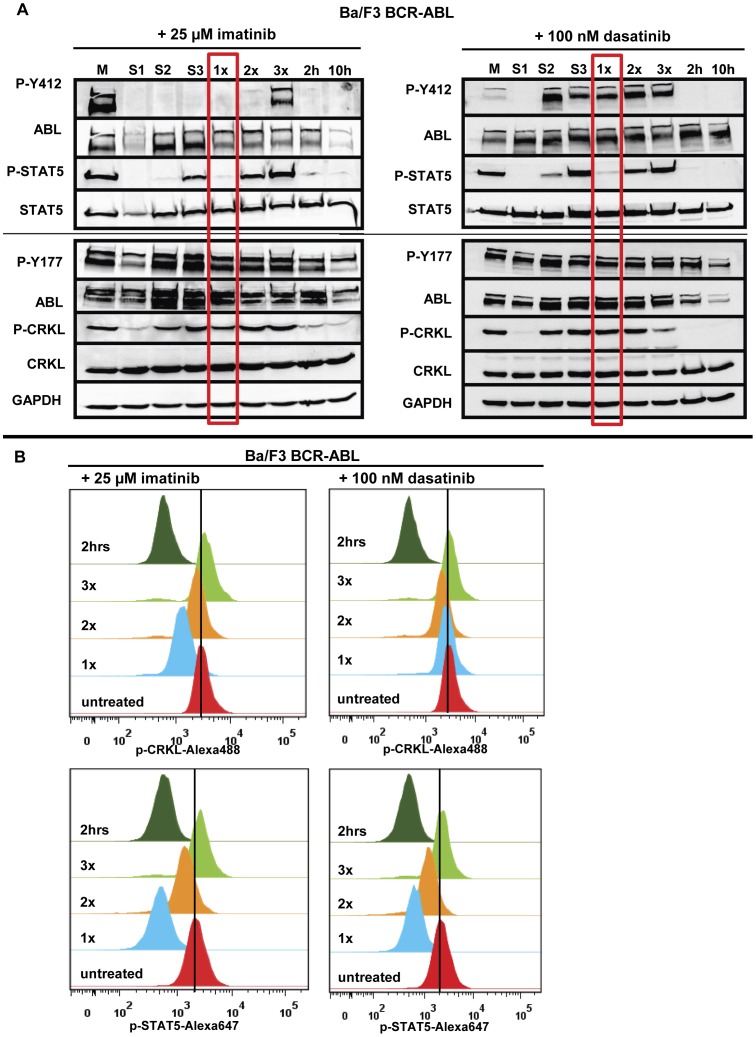
Figure 4. Recovery kinetics of intracellular signaling pathways upon HD-TKI pulse-exposure.
(A) Ba/F3-BCR-ABL cells were treated either with 25 µM imatinib or with 100 nM dasatinib for 2 hours followed by drug wash-out and consecutive replating as described in Figure 1B . Untreated cells served as positive controls for phosphorylation signals (“M”). Cells treated continuously for 2 hours or 10 hours (2 h and 10 h) served as positive controls for dephosphorylation of intracellular signalling pathways. “M”: medium; “1x”, “2x”, “3x”: Cells were treated with HD-TKI for 2 h and drug wash-out was performed 1–3x in 2 h intervals, respectively. Lysates were prepared 2 h after drug wash-out, allowing for recovery of phosphorylation signals in case of absent TKI activity. “S1”, “S2”, “S3”: Lysates were prepared upon incubation of previously untreated cells for 2 h with the respective cell culture supernatants obtained during each washing procedure. (B) FACS analysis of Ba/F3-BCR-ABL cells after HD-TKI exposure followed by repetitive drug wash-out (“1x”, “2x”, “3x”) using phosphorylation-specific antibodies against CRKL (upper panel) and STAT5 (lower panel). Cells were treated with respective TKI for 2 h as indicated and washed as explained in Figure 1B . Two hours after each washing step cells were fixed and permeabilized. Antibody staining was performed in parallel with p-CRKL-Alexa488 and p-STAT5-Alexa647 antibody. Untreated cells served as a control for phosphorylation signals. All experiments were performed at least in triplicate and one representative experiment is shown.

