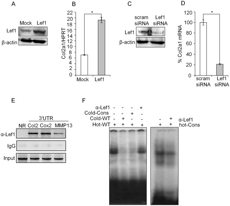
Figure 3. Lef1 dependent Col2a1 expression in primary chondrocytes.
(A, B). Primary chondrocytes were transfected with Lef1 expression plasmid or empty vector control (mock). Lef1 over-expression was confirmed by immunoblotting with antibodies against Lef1 and β-actin (control) and relative level of Col2a1 transcript was detected by qRT-PCR and expressed relative to the level of housekeeping control HPRT. (C, D) Primary chondrocytes were transfected with scrambled siRNA (mock) or Lef1 siRNA and Lef1 knock-down was confirmed by immunoblotting and relative level of Col2a1 transcript was detected by qRT-PCR and expressed relative to the level of housekeeping control HPRT. Knock-down efficiency was expressed as percentage of scrambled siRNA (control) transfected sample. (E) Physiological binding of Lef1 to the predicted conserved Lef1-binding site in the 3′ UTR was assessed by ChIP assay. Crosslinked and fragmented DNA from primary chondrocytes were immunoprecipitated with Lef1 antibody and IgG (control). PCR analysis was performed using the primers for the 3′ UTR region of Col2a1 locus. The same precipitate was also probed with primers specific for the 3′ UTR regions of Cox2 and MMP13 (positive controls) [27], [28], [29], [30] or primers specific for the non-conserved region (+16801/+17024) in the Col2a1 locus (negative control). All data are representative of three independent experiments. *P<0.05, **P<0.01. (F) EMSA was performed by incubating nuclear extract prepared from P0 stage chondrocyte with the indicated labeled probes, (hot-wild type (hot-WT) and hot-consensus (hot-Cons) and non-labeled competitor oligonucleotides (cold-wild type (cold-WT) and cold-Consensus (cold-Cons) or α-Lef1 antibody.

