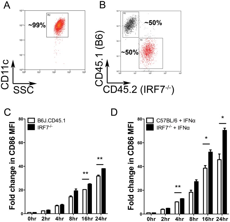
Figure 3. Exaggerated CD86 expression after TLR2 stimulation occurs in the presence of IRF7-sufficient cDCs or exogenous IFNα.
A. Representative purity of splenic CD11chi cDCs sorted from C57BL/6, B6.Irf7 −/− and congenic B6J.CD45.1 mice. B. Representative staining of cDCs from B6.Irf7 −/− and B6J.CD45.1 mice after co-culture at ∼50∶50 ratio in the presence of 10 µg/ml PAM3CSK4. C. The fold increase in expression of CD86 after TLR2 stimulation for cDCs from either strain was determined by flow cytometry at the indicated times post-stimulation. D. C57BL/6 or B6.Irf7 −/− cDCs were cultured in the presence of 10 µg/ml PAM3CSK4 and 1000 U/ml IFNα. At the indicated times post- stimulation, cells were removed and assessed by flow cytometry expression of CD86. Data are presented as mean fold increase ± SEM in surface expression of CD86 relative to unstimulated cDCs from the same strain. Data are from two experiments. * = p<0.05 ** = p<0.01, *** = p<0.001.

