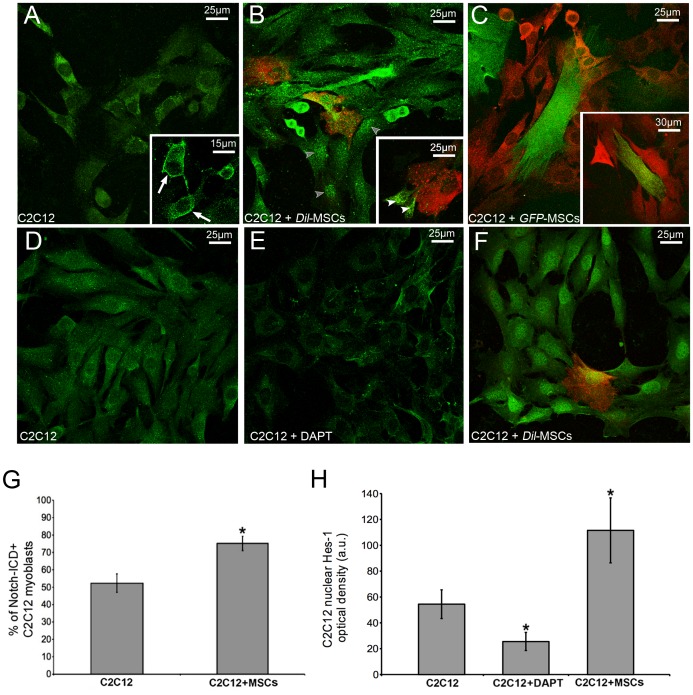
Figure 5. Notch-1 signaling is activated in C2C12 myoblasts upon co-culture with MSCs.
Confocal immunofluorescence analysis of Notch-1 (A-C) and Hes-1 (D-F) expression in C2C12 cells in single and co-culture with Dil(red)- or GFP(green)-labeled MSCs for 24 h. After the co-culture, C2C12 cells reveal a stronger reactivity for the activated Notch-intracellular domain (Notch-ICD) and for Hes-1, which is visible inside the nucleus. As shown in the inserts, Notch-1 is preferentially located at the cell membrane (arrows) in the single cultured C2C12 cells, whereas it is found within the cytoplasm (white arrowheads) and nucleus (grey arrowheads) in the co-cultured cells. E) C2C12 myoblast were treated with 5 µM DAPT to inhibit Notch-1 activation and assayed for Hes-1 expression. The corresponding quantitative analysis is reported in the histograms (G,H). Data represent the results of at least three independent experiments with similar results. *p<0.05.

