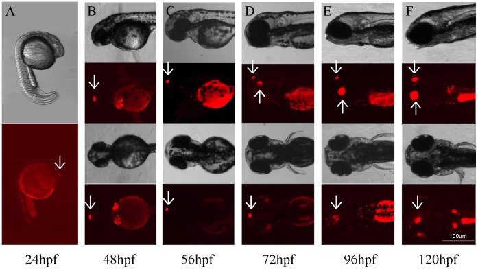
Figure 1. Bright-field and fluorescent images of developing embryos at different developmental stages that show Tg(Gnat2:gal4-VP16/UAS:nfsB-mCherry) transgene expression in the pineal gland and retina.
(A) At 24 hpf, mCherry was detected in the pineal gland (arrow). (B) Strong mCherry expression was seen in the pineal gland (arrows). (C–F) Between 56 and 120 hpf, mCherry was detected in both the pineal gland (downward arrows) and retina (upward arrows). The intensity of transgene expression increased during embryonic development.

