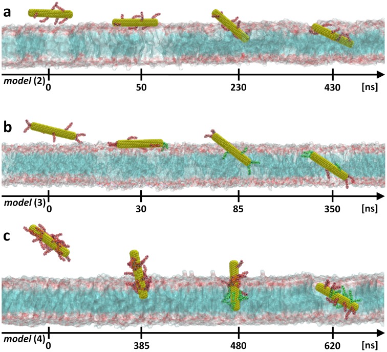
Figure 4. Uptake path of closed f-CNTs.
Results obtained from unconstrained MD simulations for: a, closed and low degree side functionalized SWNT [model (2)]; b, low degree side and tip functionalized SWNT [model (3)]; c, or highly side functionalized SWNT [model (4)]. Note that f-CNTs can be completely taken up only when the cationic functional groups are deprotonated (cf. text). The yellow surface represents the SWNT core while the amino functional groups attached to the latter are shown as red (charged form) or green (neutral form) atoms. The lipid membrane head and tails sections are shown as pale red and blue surfaces, respectively. For clarity, water molecules and counterions are not shown.

