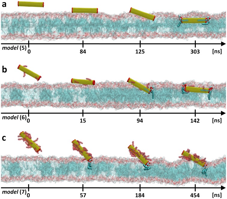
Figure 6. Uptake path of open ended f-CNTs.
Results obtained from unconstrained MD simulations with: a, opened and non functionalized SWNT [model (5)]; b, low degree functionalized and opened SWNT [model (6)]; c, or highly functionalized and opened SWNT [model (7)]. The yellow surface represents the SWNT core. H atoms (red balls) are used to passivate the SWNT edges. The TEG-NH3 + functional groups (red surfaces) are attached to the SWNT surface. The lipid membrane head and tails sections are shown as red and blue surfaces, respectively. Note that at the end of each trajectory, a single lipid molecule stays strongly anchored at the SWNT tips. This anchored lipid molecule is shown explicitly as blue (acyl chains), red (carboxyl group) and blue/white (phosphatidylcholine headgroup) balls. For clarity reasons, water molecules and counterions are not shown.

