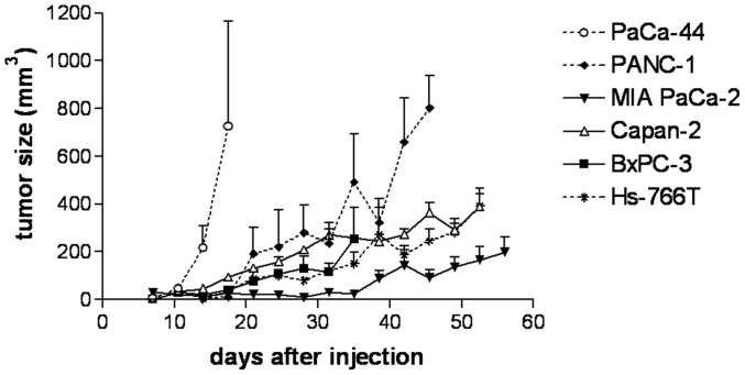
Figure 1. Tumor growth development of subcutaneously growing tumors.
5×10⁶ cells of each pancreatic cell line were subcutaneously injected into the left flank of C.B-17/IcrHsd-Prkcdscid Lystbg mice at day 0. Visible tumors were measured twice a week with a caliper and size was calculated by the formula “length×width×width/2”.

