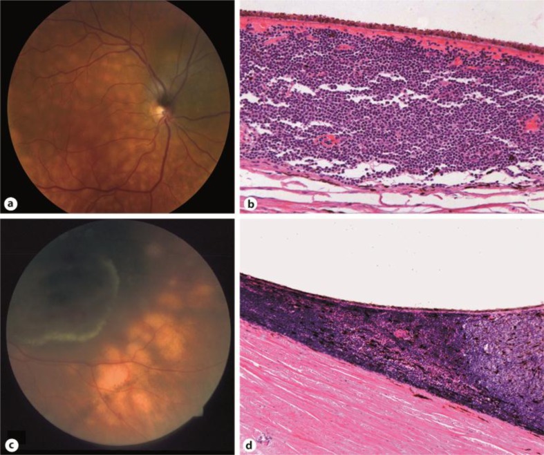
Fig. 1.
a Fundus of patient 1 showing a juxtapapillary choroidal melanoma surrounded by creamy yellow choroidal spots. b Patient 1. Dense lymphocytic choroidal infiltrates outside the melanoma area. The lymphocytes are diffusely, but tightly packed unlike inflammatory lymphocytic infiltrate and show irregular nuclei and nuclear membranes. Paraffin section. Original magnification: 200×. c Characteristic yellow spotted fundus of patient 2 with choroidal melanoma in inferior temporal area. d Patient 2. Dense lymphocytic infiltrate at the margins of the melanoma extending towards the equator. Celloidin section. Original magnification: 100×.

