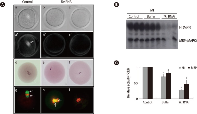
Figure 5.
Changes in spindle, chromosome, and kinase activity in oocytes after Tkt RNAi. (A) Spindle structure of control MI (a) and arrested MI oocytes (b, c) was observed non-invasively following microinjection of Tkt dsRNA using a Pol-Scope. Upper column: bright fields; lower column: dark fields. Chromosome status was evaluated by Orcein staining (d-f). Bars indicate 25 µm. The abnormalities in the spindles and chromosomes in the Tkt RNAi oocytes (h, i) were confirmed by immunofluorescence staining. Green, tubulin; red, DNA. Arrows indicate spindles. (B) The amount of substrate phosphorylation following Tkt RNAi treatment. Phosphorylation of the substrates H1 and MBP reflects the kinase activities of MPF and MAPK, respectively. Each lane contains one oocyte of the indicated group. (C) The amount of phosphorylated H1 and MBP, namely the relative activity of each kinase, was calculated by quantifying phosphorylation of the substrates. The experiments were repeated at least three times, and the data are presented as the mean±SE (ap<0.05). Tkt, transketolase; MPF, maturation promoting factor; MAPK, mitogen-activated protein kinase; H1, histone H1; MBP, myelin basic protein; MI, metaphase I.

