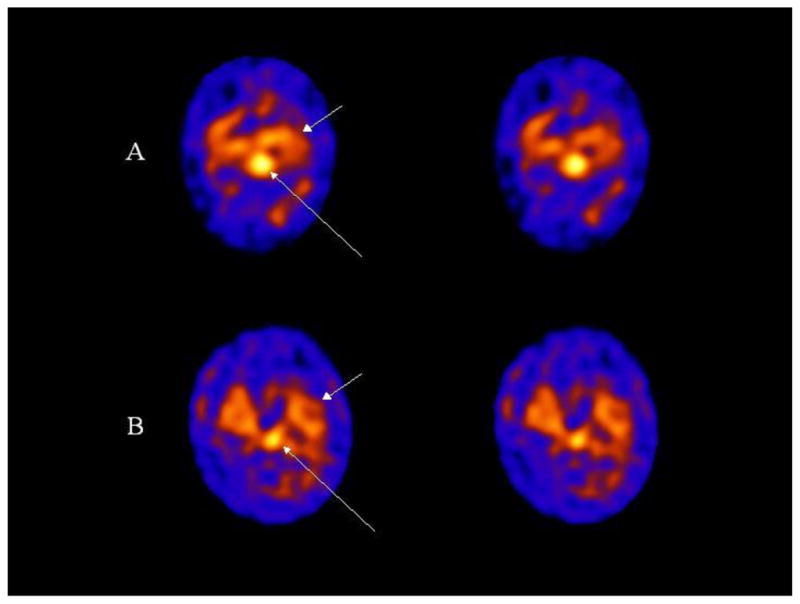
Figure 1.

Transaxial 123I ADAM SPECT images are shown of a control (A) and depression (B) subject. The long arrows point to uptake in the midbrain which is markedly decreased in the subject with depression compared to the control subject. Non-specific binding in the cerebellum is observed below the midbrain uptake on the images (posteriorly). The short arrows point to 123I ADAM binding in the medial temporal lobe.
