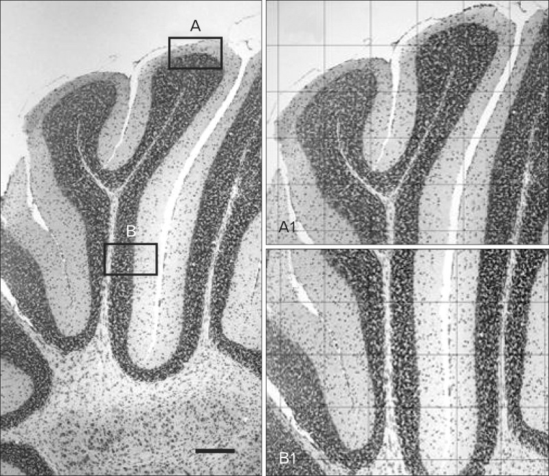
Fig. 1.
A representative coronal section stained with cresyl violet of cerebellum at control Balb/c mice (P18) group. The left picture shows a simple lobule of cerebellum in 40× magnification. A1, apex of lobule in 100× magnification (grid: 200 µm×200 µm); B1, depth of lobule in 100× magnification (grid: 200 µm×200 µm). P, postnatal day. Scale bar=200 µm.

