
Figure 3. N-acetyl-cysteine (NAC) partially rescues reduced glutathione (GSH)-depleted U-87 MG cells.
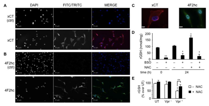
U-87 MG astroglioma cells possess the cystine/glutamate antiporter system, composed of a light subunit, catalytic unit (xCT) (A) and a regulatory heavy chain (4F2hc) (B), both of which localize predominantly at the plasma membrane. Astroglioma cells abundantly synthesized both subunits for continuous uptake of cystine in exchange for intracellular glutamate, thus giving rise to a clear intracellular staining pattern. The first column shows stained nuclei (DAPI), whereas the second column represents the TRITC/red (for xCT) or FITC/green (for 4F2hc) channel. These two channels are shown in black and white and in color when the results from the two channels are merged in the third column (magnification 20×). (C) Detailed images of the two subunits in U-87 MG cells are shown at magnification 100×, which revealed dispersed intracellular presence as well as presence at the plasma membrane. Scale bars = 10 μm. (D) The xc system is functional in U-87 MG. Cells were cultured in the absence or presence of known compounds capable of either decreasing (buthionine sulfoximine, BSO) or increasing (N-acetyl-cysteine, NAC) the intracellular concentration of reduced glutathione (GSH). Treatment with BSO (100 μM) induced an approximate tenfold decrease in GSH concentration. Cells treated for 24 h with NAC (5 mM) displayed a 70% to 80% increase in the level of intracellular GSH. On the other hand, the level of intracellular GSH in cells pretreated with BSO and subsequently with NAC was not rescued due to the irreversibility of the action of BSO on γ-glutamyl-cysteine synthetase. *P values < 0.05; **P value < 0.001 compared with untreated cells (Student paired t test). (E) U-87 MG cells treated with Vpr-deficient or Vpr-containing conditioned medium displayed an increase in intracellular GSH concentrations. U-87 MG cells exposed to extracellular Vpr-containing conditioned medium and treated with NAC for 24 h partially recovered cells from the induction of oxidative stress. *P value < 0.001 of untreated compared to Vpr-treated U-87 MG. **P value < 0.05 compared to U-87 MG exposed to Vpr-containing conditioned medium in the absence of NAC (Student paired t test). P-values are determined as percent over control at 0 h time point.