
Figure 4. Extracellular rVpr induces a significant decline in intracellular ATP, reduced GSH, GSH/GSSG ratio, and accumulation of ROS in U-87 MG cells.
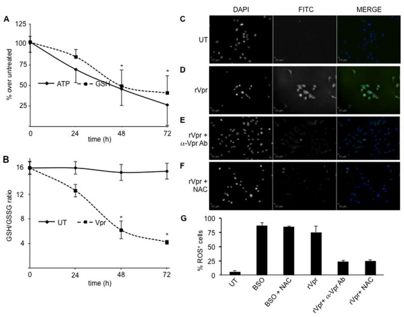
(A) U-87 MG astroglioma cells exposed to 5 μg/ml of extracellular recombinant Vpr showed a time-dependent decrease in ATP levels up to fourfold compared to untreated cells after 72 h exposure (black line). In a similar time-dependent manner, the concentration of GSH decreased about 2.5-fold after 72 h (dotted line). *P values < 0.05 compared to untreated cells (Student paired t test). (B) The intracellular levels of reduced and oxidized glutathione and the GSH/GSSG ratios were determined after incubation of U-87 MG astroglioma cells in either the absence or the presence of 5 μg/ml of recombinant Vpr for the indicated periods of time. Whereas untreated cells maintained constant levels of reduced and oxidized glutathione (GSH/GSSG stable), Vpr-treated cells exhibited a time-dependent decrease in the GSH/GSSG ratio. While untreated U-87 MG cells displayed no staining with the ROS-sensitive RedoxSensor Red CC-1 dye (C), exposure to extracellular recombinant Vpr (5 μg/ml) induced abundant accumulation of ROS (D), which was partially blocked by treatment with either anti-Vpr antibody (E) or addition of 5 mM NAC (F) for 24 h. In all images, the first column shows stained nuclei (DAPI), whereas the second column represents the FITC/green (RedoxSensor Red CC-1 dye) channel. These two channels are shown in black and white and in color when the results from the two channels are merged in the third column. All magnifications are 20×. Scale bars = 10 μm.