
Figure 1.
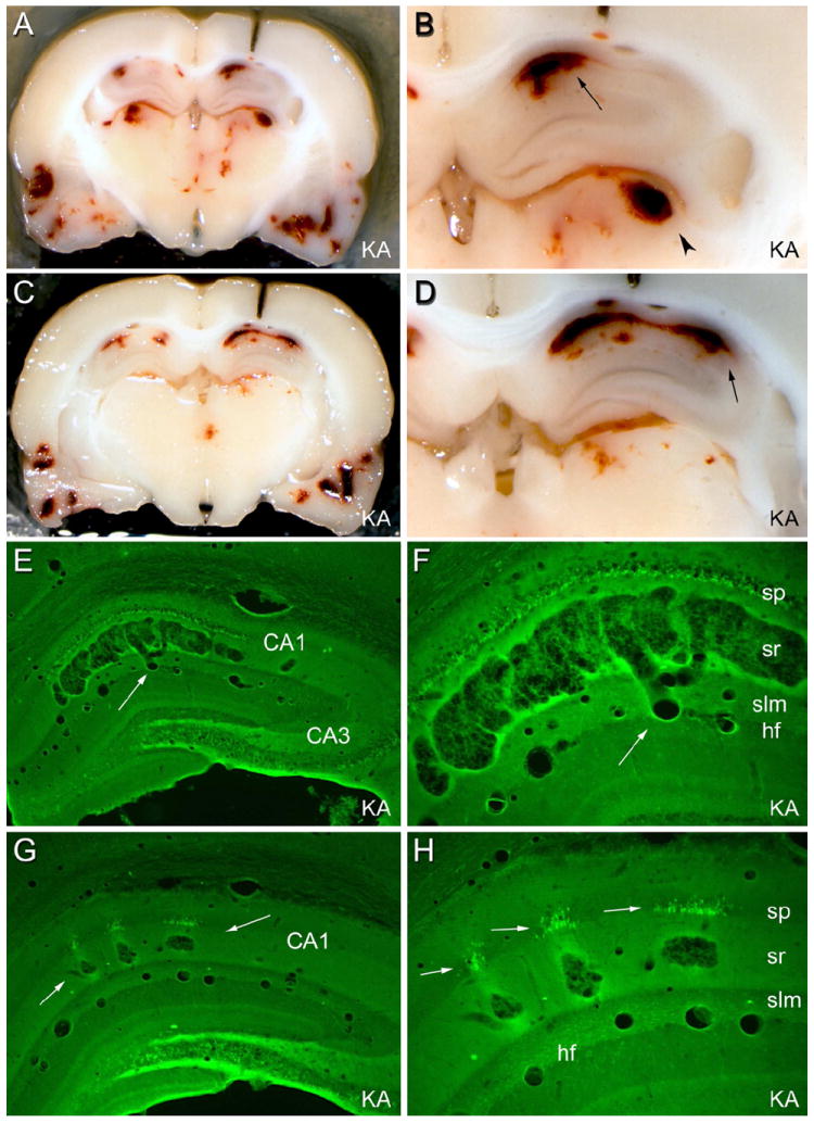
Brain structure 3 days after status epilepticus induced by systemic injection of kainic acid (KA; 12 mg/kg sc). In some animals given kainate or pilocarpine systemically, prolonged status epilepticus caused apparent hemorrhages in a multitude of brain structures, which were not cleared by vascular perfusion. (A) and (C): two coronal views of the same brain during the sectioning process. Note apparent hemorrhagic foci in the hippocampi, thalamus, and temporal cortices. (B) and (D): foci preferentially involve the CA1 pyramidal cell layer (arrow) and the dorsolateral thalamus (arrowhead). (E) and (F): in a Fluoro Jade B-stained section from the brain shown in (A)-(D), degenerating neurons are fluorescent. Note that the area CA1 pathology consists of a vascular expansion that is continuous with a capillary in the hippocampal fissure (hf; arrow), not an extravascular hemorrhage. (G): in a different kainate-treated rat, smaller focal vascular expansions occurred within the stratum radiatum (arrows). (H) At higher magnification, degenerating CA1 pyramidal cell somata were evident only adjacent to the vascular pathology. These results suggest that, in some cases, CA1 pyramidal cell layer injury in rats subjected to prolonged status epilepticus may be ischemic in nature, rather than excitotoxic. Abbreviations: sp: stratum pyramidale; sr: stratum radiatum; slm: stratum lacunosum-moleculare; hf: hippocampal fissure. Magnifications: 5X (A and C); 13.5X (B and D); 22X (E and G); 55X (F and H). From Sloviter, 2005.