
Figure 2.
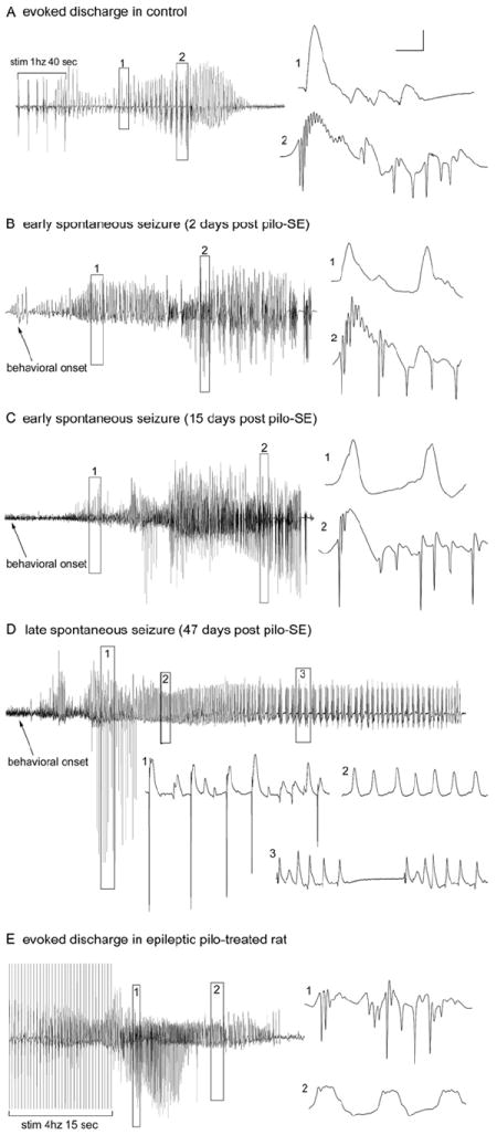
Evoked and spontaneous granule cell activity recorded from the granule cell layers of chronically epileptic, pilocarpine treated-rats. A: Granule cell layer seizure discharges evoked in a control animal (60 days post-saline treatment) by perforant path stimulation at 1 Hz (with paired-pulses 40 msec apart) for 40 sec. Note that evoked granule cell activity generated both low-frequency, positive-going field potentials (expanded box 1) and high-frequency, negative-going population spikes (expanded box 2). B: In an epileptic animal 2 days post-SE, spontaneous granule cell layer activity recorded during a spontaneous behavioral seizure exhibited both positive-going field potentials (expanded box 1) and negative-going epileptiform discharges (expanded box 2) that were qualitatively similar to those evoked in the control rat in A. C: Thirteen days later, spontaneous granule cell layer activity still exhibited high-frequency, negative-going epileptiform discharges. D: Forty-seven days after SE, the spontaneous granule cell layer activity recorded in the same animal during a spontaneous seizure exhibited single population spikes (expanded box 1) and positive-going field potentials (expanded boxes 2 and 3) but no seizure discharges (repetitive population spikes). Note that granule cells generated only single population spikes ~7 seconds after the onset of the observed behavioral seizure (expanded box 1) and then generated only field potentials for the duration of the behavioral seizure. E: Evoked granule cell seizure afterdischarges in the same chronically epileptic, pilocarpine-treated rat 45 days post-SE (tested 2 days prior to the activity shown in D), illustrating that, despite the lack of spontaneous seizure discharges in the chronic epileptic state, abnormally high-frequency (4 –10Hz) stimulation could evoke granule cell seizure discharges (expanded box 1), as well as positive-going field potentials (expanded box 2). The absence of seizure discharges recorded from the granule cell layers during spontaneous behavioral seizures was a consistent observation in epileptic animals subsequently shown to have dentate hilar cell loss and mossy fiber sprouting. Calibration bars: 3.5 mV, 30 msec in A; 5 mV, 2 sec for B; 4 mV, 30 msec for B1,2; 2.5 mV, 2 sec for C; 3 mV, 100 msec for C1–3; 3 mV, 2 sec for D; 4 mV, 25 msec for D1,2. From Harvey and Sloviter, 2005.