
Figure 4.
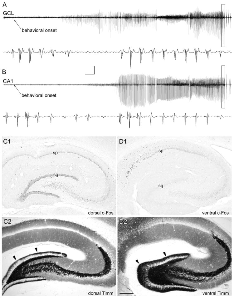
Atypical recruitment of granule cell and CA1 pyramidal cell discharges during a spontaneous seizure in a chronically epileptic rat. A,B: In this animal, perfusion fixed 56 days post-SE, seizure discharges were recorded from both dorsal hippocampal granule cell (GCL) and CA1 pyramidal cell layers beginning approximately 25 seconds after the behavioral seizure onset. C1: c-Fos immunostaining of the dorsal hippocampus in a coronal section revealed c-Fos expression in all hippocampal neurons. D1: c-Fos immunostaining of the ventral hippocampus in a horizontal section from the same rat revealed c-Fos expression in pyramidal layer neurons but not in the dentate gyrus (sg; stratum granulosum). C2,D2: Note that Timm staining revealed less extensive mossy fiber sprouting in the dorsal hippocampus (C2) than in the ventral hippocampus (D2), and a lack of c-Fos expression in the more heavily mossy fiber-sprouted ventral dentate gyrus. Calibration bars: 10 mV, 5 seconds (compressed areas); 6 mV, 75 msec (expanded boxes). Scale bar: 400 um in D (applies to C,D). From Harvey and Sloviter, 2005.