
Figure 6.
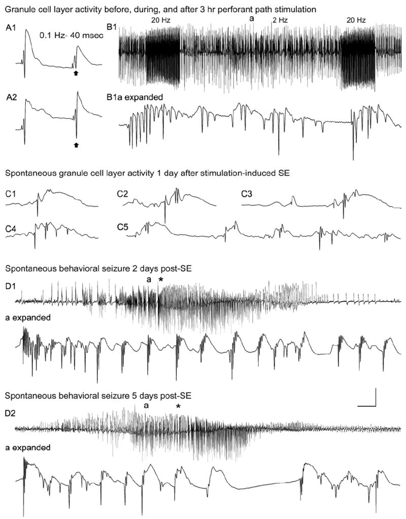
Dentate granule cell excitability and spontaneous activity before and 1–5 days after 3 hours of perforant pathway stimulation-induced convulsive status epilepticus (SE). A1: Before SE, paired-pulse perforant pathway stimulation at 0.1 Hz and an interstimulus interval of 40 msec evokes granule cell responses that exhibit partial suppression of the amplitude of the second population spike (arrow). A2: Three days after 3 hours of SE, the identical afferent stimulation failed to suppress the second population spike (arrow). B1: Granule cell layer activity during 3 hours of perforant pathway stimulation in the same awake rat. The stimulation paradigm involved continuous stimulation at 2 Hz with paired pulses delivered at a 40-msec inter- pulse interval plus 10-second-long 20-Hz trains delivered once per minute. Note the morphology of the granule cell epileptiform discharges during the 2 Hz intertrain interval (a) in the expanded trace (B1a expanded). C: On the first day after 3 hours of stimulation- induced SE, a granule cell layer electrode recorded spontaneous granule cell field “EPSPs” and population spikes that closely resemble the evoked responses in A. D: Granule cell layer activity during spontaneous behavioral seizures during the first week post-SE. D1: On day 2 post-SE, granule cell layer activity amplitude increased before the behavioral onset of the second behavioral seizure on that day (marked by asterisk). D1a (expanded): Expanded trace of the region above marked “a,” showing that the high-amplitude activity in D1 consisted of granule cell epileptiform discharges that preceded the behavioral seizure onset (asterisk). D2: Three days later, the fourth spontaneous behavioral seizure exhibited nearly identical features, including high-frequency granule cell epileptiform discharges (D2a expanded) that preceded the behavioral seizure-onset (asterisk). Calibration bars: 14 msec and 9mV in A; 7 seconds and 9 mV in B1; 46 msec and 9mV in B1 (expanded); 40 msec and 9 mV in C; 3.4 seconds and 9 mV in D1,D2; 53 msec and 9 mV in D1 (expanded); 60 msec and 9 mV in D2 (expanded). From Bumanglag and Sloviter, 2008.