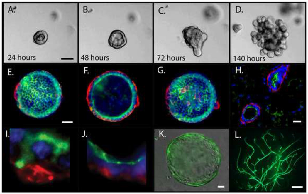
Figure 2. Organoid 3D culture and mammary reconstitution.
Primary MECs grown in EHS matrix develop into organoids that retain the cellular organization, polarity and differentiation capacity of the mammary gland. A-D) Aggregated primary MECs were embedded in EHS matrix and cultured for 140 hours. A and B) Organoids organize into hollow cysts by 24-48 hours. C) Cysts collapse and begin branching around 72 hours in culture. D) By 140 hours the cyst is fully branched. E-G) Immunofluorescence of organoids that show the cellular organization of myoepithelial cells (Keratin 14, red) and luminal cells (Keratin 18, green). E) Top F) middle and G) bottom of cyst. H) Mouse mammary gland immunofluorescence of myoepithelial cells (smooth-muscle α-actin, red) and luminal cells (mucin, green). I) High magnification of Keratin 14 (red) and Keratin 18 (green) cells in a cyst growing in EHS matrix showing the distinct myoepithelial and luminal cell layers. J) Magnified image of a cyst growing in EHS matrix demonstrating polarity of luminal cells with smooth-muscle α-actin in red and the apical marker ZO1 in green. K-L) Viral-mediated genetic modification of MEC's in culture and a mammary outgrowth. K) A cyst grown in EHS matrix derived from a MEC transduced with a GFP expressing lentivirus. L) Mammary outgrowth derived from MECs transduced with a GFP expressing lentivirus. A-D) Scale bar=50um E-G) Scale bar=20um H) Scale bar=25um K-L) Scale bar=1mm

