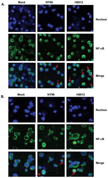
Fig. 5.
Nuclear translocation of NF-κB in WNV-infected macrophages and kidney epithelial cells. Immunofluorescence photomicrographs of mouse macrophage cells (A) and kidney epithelial cells (B) infected with mock, WNV NY99 and WNV H8912 (MOI =1). Mock-infected and WNV NY99 or H8912-post-infected cells were double labeled on day 3 post-infection with NFκB-p65 antibody (Green signal) and Hoechst dye for nuclear counterstaining (blue signal). Arrow points to cells in which NF-κB translocation to the nucleus was detected.

