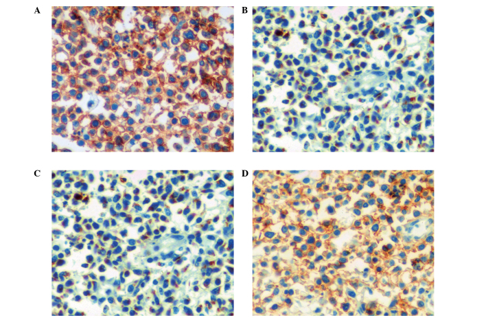
Figure 4.
Immunohistochemical stain demonstrating (A) LCA-positive tumor cells completely surrounding and infiltrating seminiferous tubule, (B) TIA-1-positive tumor cells completely filling the entire field of vision, (C) CD43-positive tumor cells completely filling the entire field of vision and (D) CD45RO-positive tumor cells completely filling the entire field of vision. (A and D) Original magnification, ×100. (B and C) Original magnification, ×400.

