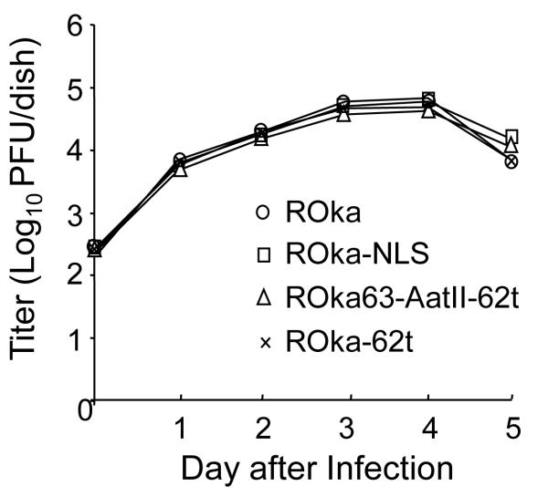
Fig. 3.
Growth of VZV ROka, ROka-NLS, ROka63-AatII-62t, and ROka-62t in cell culture. Melanoma cells were infected with each virus and at days 1 to 5 after infection the cells were treated with trypsin and the titer of cell-associated virus was determined on melanoma cells. Each data point shown is the average of two separate values.

