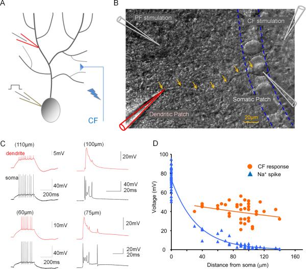
Figure 1.
Depolarization-evoked Na+ spikes and CF responses in somato-dendritic double-patch recordings. (A) Recording configuration. Patch-clamp recordings were obtained from the soma and the dendrite of Purkinje cells. Depolarizing current pulses were applied through the somatic patch electrode. The CF input was activated using extracellular stimulation with a glass pipette filled with ACSF. (B) DIC image illustrating the double-patch configuration. Arrows outline the course of the primary dendrite. Glass pipettes for PF and CF stimulation, respectively, are shown in the upper left and right corners. (C) Examples of Na+ spikes evoked by somatic depolarization (left) and CF responses (right) recorded at dendritic locations (red traces) and the soma (black traces). (D) Distance dependence of Na+ spike (blue) and CF response (orange) amplitudes. The fitted lines were obtained using the least-square method.

