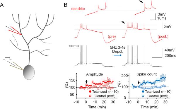
Figure 3.
Repeated somatic injection of depolarizing currents enhances the frequency and dendritic amplitude of Na+ spikes. (A) Recording configuration. Na+ spikes evoked by single depolarizing current pulses injected into the soma were recorded in the test periods before and after repeated somatic current injection at 5Hz. (B) The depolarization protocol enhances the number of action potentials and the amplitude of dendritic Na+ spikes (red traces; enlarged in the insets). Lower left: Time graph showing Na+ spike amplitude changes recorded in the dendrite after tetanization (closed dots; n=10) and under control conditions (open dots; n=5). Lower right: Time graph showing associated changes in the spike count after tetanization (closed dots; n=10) and under control conditions (open dots; n=5). Arrows indicate tetanization. Error bars indicate SEM.

